


Hip Dysplasia and Hip Dislocation
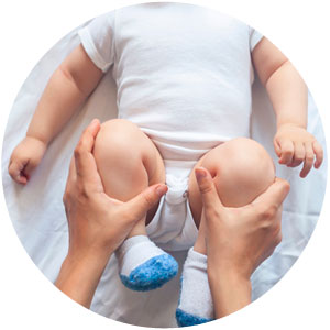
Developmental dysplasia of the hip is characterized by insufficient development of the hip socket, resulting in insufficient coverage of the ball of the hip. This results in instability or dislocation of the hip.
Hip dysplasia is detected with ultrasonography, which is performed during the first 2 weeks after birth and at 6 to 8 weeks after birth. Almost all cases can be treated with a harness that needs to be used day and night until ultrasound confirms sufficient correction.
Some cases of hip dislocation require casting to center the hip within the socket before harness treatment is begun. If the hip cannot be reduced but remains dislocated, open surgical reduction is necessary.
Residual dysplasia, with the hip within the socket but with insufficient coverage and instability, can result from insufficient correction with the harness or from mild dysplasia missed during the initial ultrasonographic examinations. Residual dysplasia can lead to arthritis and pain in adulthood. If diagnosed early, it can be successfully treated by performing pelvic redirecting osteotomy (improving the coverage of the hip through surgical correction of the position of the hip socket).
Please note that I am not performing hip screening ultrasonography for newborns in my office. If hip dislocation has been diagnosed, I can perform treatment and surgical correction.
Transient Synovitis of the Hip
Transient synovitis of the hip is an inflammation in the hip joint that causes pain. It mostly affects children between the ages of 4 and 11 years and often occurs after viral respiratory infection. Children complain about pain in the hip that might radiate to the knee and lower limb.
Most cases resolve well with rest and anti-inflammatory medication. However, it is important to rule out other causes, especially septic arthritis of the hip caused by bacteria. Septic arthritis is rare but requires specific diagnosis and urgent treatment.
Slipped Capital Femoral Epiphysis (SCFE)
SCFE affects the hip at the growth plate. The epiphysis (top part of the hip ball) slips at the growth plate from the upper part of the remainder of the femur (thigh bone). The growth plate is soft and is therefore a weak spot until it closes at the end of growth. SCFE can occur with trauma, sports, or obesity.
SCFE can happen acutely with trauma, characterized by instant pain and limping. However, SCFE can also occur slowly with a small amount of pain that can radiate and is often described as knee pain. As a result, diagnosis can be delayed or missed.
The most common type is called acute on chronic. This type is characterized by mild pain over a prolonged period (the growth plate gets wider and unstable) and then a sudden increase of pain and limping (the actual slip).
After the diagnosis is confirmed, surgery is necessary to stabilize the slipped epiphysis and to prevent further slipping and deformity. This is usually done by pinning the hip across the physis (growth plate). In severe cases, the hip needs to be taken out of the socket to put the epiphysis back in place before stabilization.
Perthes Disease (Legg-Calve-Perthes Disease)
Perthes disease is usually seen in children who are 4 to 9 years old. The blood supply to the hip ball is temporarily interrupted, which leads to avascular necrosis (death of tissue) of the hip. The children complain of pain, and the range of motion of the hip becomes limited. The true reasons for and the mechanisms of Perthes disease are not clear.
The disorder has typical stages during which the hip partially collapses to restore afterward. However, the hip might end up being deformed and the relation of the hip ball to the hip socket might be different. This can lead to early hip pain and arthritis. Generally, the prognosis is better for younger children.
Early diagnosis and treatment are important. During the early stage, infusion with special medication that increases blood flow is possible. Reducing the mechanical stress and loading of the hip is the cornerstone of further treatment. Although normal activity can be continued, impact sports, running, and jumping are discouraged. Physical therapy to preserve or restore range of motion is recommended. For some children, surgery to increase and improve the coverage (containment) of the hip can improve the outcome.